Main technical parameters:
1. The nominal value of the viewing angle is 12°, and the tolerance is ±10°
2. The field of view is 100°, and its limit deviation is ±15%.
3. Resolution: At the working distance (10mm), the resolution should be ≥7.0 lp/mm.
4. The depth of field of observation should not be less than 2mm~50mm.
5. Model corresponding specifications and dimensions

Rigid electronic ureteroscope
The mirror body has high rigidity and low deflection
The structure is strong while allowing the mirror body to bend slightly and rebound quickly.
Sapphire lens, integrated mirror body
The front objective lens is sealed with a sapphire lens, which is not easy to wear and prolongs its life. The laser welding seals the mirror body to ensure that it will not fog or get water in during surgery or disinfection.
High-definition image quality and wide surgical field of view
160,000 pixels, the clarity is more than 5 times that of traditional ureteroscopes. 100° wide field of view, 12° viewing angle.
The depth of field range is 2-100mm, and the ultra-large field of view brings a wide field of view to meet the needs of clear observation of the surgical site.
Automatic elimination of moiré patterns
Accurately eliminate moiré patterns, no interference in the picture, and improve image quality.
Image automatic return function
The lens has a built-in position sensor, and the 360° rotating mirror can still keep the field of view always forward, making surgical observation more flexible.
Hand-eye coordination helps make surgery safer and more efficient
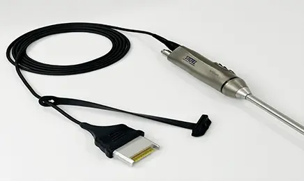
Transfer-free integrated design
The image light source integrated connector eliminates the need for external light guides and cameras, simplifies operation and reduces operator fatigue.
One-handed operation is comfortable to hold
The mirror body is ergonomically designed and weighs less than 315g, which is half the weight of traditional camera optical endoscopes. It saves effort when operated with one hand and improves the stability of holding the mirror.

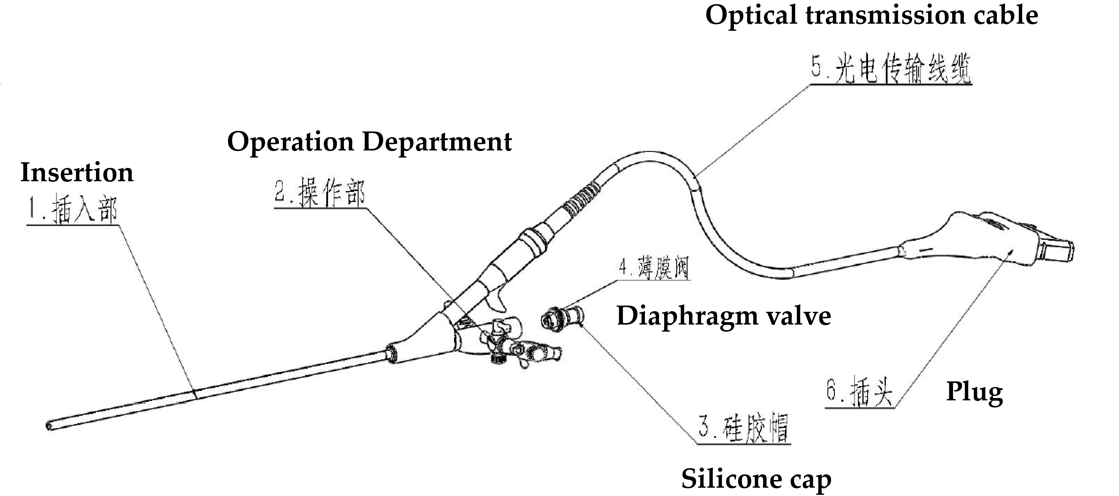
(1) Insertion part Insert into human body
(2) Operation part Operate rigid electronic cystoscope
(3) Silicone cap Used to seal the instrument channel tube during product cleaning, disinfection and sterilization
(4) Membrane valve Used to fix and seal the instrument during use with matching accessories to prevent shaking and leakage
(5) Photoelectric transmission cable Used to realize electrical connection between electronic endoscope image processor device and endoscope
(6) Plug Electrically connect with the matching electronic endoscope image processor interface and fix the photoelectric transmission cable
_-_副本.webp)


